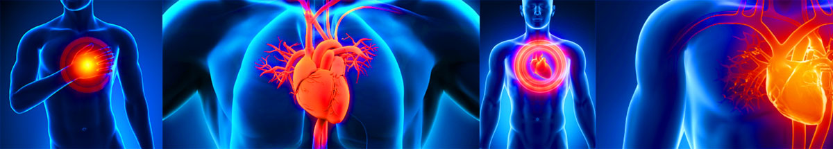
If you happen to be experiencing chest discomfort, breathlessness, dizziness, palpitations or other worrying symptoms, and you’re looking for a team of highly dedicated cardiac professionals to address your concerns then our experienced and friendly staff will quickly and efficiently provide a comprehensive checkup to make sure you’re not in harm’s way.
With offices at several locations on the Gold Coast and now also in Lismore, we work closely with your GP to ensure you access the best and safest treatment possible.
These conveniently co-located highly credentialed independent cardiologists together with CCA’s team of cardiac sonographers and technicians, and our administrative staff, will take care of every detail.
In our hands peace of mind is part of the deal.
Symptoms & Treatments
Heart Attack
The coronary arteries supply blood to the heart muscle. When the coronary arteries become narrow or blocked, blood flow to the heart is reduced. This decrease in blood flow to the heart deprives the heart muscle of oxygen.
Heart attack (also called myocardial infarction) is when part of the heart muscle is damaged or dies because it isn’t receiving oxygen. Most heart attacks are caused by a blockage in the coronary arteries.
If you suspect symptoms of heart attack, you should call for an ambulance or seek immediate medical attention.
The main function of the heart is to deliver the oxygen-rich blood to every cell in the body.
The arteries are the passageways through which the blood is delivered and the veins are the passageways through which the blood is collected and returned to the heart.
The coronary arteries supply blood to the heart muscle. When the coronary arteries become narrow or blocked, blood flow to the heart is reduced. This decrease in blood flow to the heart deprives the heart muscle of oxygen.
Heart attack (also called myocardial infarction) is when part of the heart muscle is damaged or dies because it isn’t receiving oxygen.
Complications
Complications depend upon the location and extent of the heart damage (due to blocked blood supply). Early intervention and treatment could prevent these complications.
Cardiac arrhythmias
Cardiac arrhythmias are disruptions in the natural rhythm of the heartbeat
Cardiac Failure
Here the heart fails to pump blood to meet the metabolic demands of the body
Pericarditis
Pericarditis is the inflammation of the pericardium, the outer covering of the heart that acts as a shock absorber for the heart
Recurrent heart attacks
Increased risk of heart attacks and angina in the future
Blood clots (Thromboembolism)
Blood clots may be formed due to irregular rhythms and prolonged immobility. You may be prescribed blood-thinning agents that need to be monitored with regular blood tests
Atherosclerosis
Atherosclerosis is a condition in which fatty material is deposited along the walls of arteries. This fatty material (often called plaque) thickens, hardens, and may eventually block the arteries.
Atherosclerosis of the coronary arteries is the most common cause of heart attack.
Risk factors
Risk factors for atherosclerosis and heart attack include
Family history
Hypertension (High Blood Pressure)
High Cholesterol or other fat levels in blood
Inactive lifestyle- Obesity/ overweight/ lack of exercise
Diabetes (High blood sugar)
Cigarette Smoking
How will you feel?
Chest pain is the most common complaint in heart attack. Unlike angina, pain does not subside on resting.
However, the symptoms may be different.
For example,
Fullness, uncomfortable pressure, squeeze in the middle of the chest
Tightness, burning or a heavy weight over your chest
Pain may radiate to your shoulders, neck, arms, upper abdomen, back or jaw
20% of the patients with heart attack have no pain. This is seen in diabetics, high blood pressure, and elderly patients.
Heart attack is a medical emergency and if you suspect symptoms of heart attack, you should call for an ambulance or seek immediate medical help.
(EKG or ECG) Electrocardiogram
EKG is a test to measure the electrical activity of the heart and provides your doctor with information about your heart rate, rhythm, size of the heart chambers and previous damage to the heart. It is non-invasive and painless and is performed by attaching electrodes to various parts of the b
Angina
Angina Pectoris (“Angina”) is a recurring pain or discomfort in the chest that happens when some part of the heart does not receive enough blood.
Angina is a common symptom of coronary artery disease that occurs when the coronary arteries are narrowed or blocked. It is usually relieved within a few minutes by resting or by taking prescribed angina medicine.
Important Note
Not all chest pain is angina and therefore should always be evaluated by a Physician.
The main function of the heart is to deliver oxygen-rich blood to every cell in the body.
The arteries are the passageways through which the blood is delivered and the veins are the passageways through which the blood is collected and returned to the heart.
The coronary arteries supply blood to the heart muscle. When the coronary arteries become narrow or blocked, blood flow to the heart is reduced. This decrease in blood flow to the heart deprives the heart muscle of oxygen. The heart responds to the lack of oxygen by sending out signs in the form of pain called angina.
Atherosclerosis
Atherosclerosis is a condition in which fatty material is deposited along the walls of arteries. This fatty material (often called plaque) thickens, hardens, and may eventually block the arteries.
Atherosclerosis of the coronary arteries is the most common cause of angina.
Risk factors
Risk factors for atherosclerosis and angina include
Family history
Hypertension (High Blood Pressure)
High cholesterol or other fat levels in blood
Inactive lifestyle- Obesity/ overweight/ lack of exercise
Diabetes (High blood sugar)
Cigarette smoking
Triggers
Physical exertion is the most common trigger for angina.
Others triggers include
Emotional stress
Extreme cold or heat
Heavy meals
Alcohol consumption and cigarette smoking
How will you feel?
Chest pain is the most common complaint in angina. However, the symptoms may be different.
For example
Fullness, uncomfortable pressure, squeezing sensation in the middle of the chest
Tightness, burning, or a heavy weight over your chest
Pain may radiate to your shoulders, neck, arms, upper abdomen, back or jaw
Important Note
Not all chest pain is angina and therefore should always be evaluated by a Physician.
Diagnoses
To diagnose angina, your doctor may use one of more of the following
Medical History
This includes Physical examination, questions about your symptoms, risk factors, personal history, and family history of any heart disease.
(EKG or ECG) Electrocardiogram
EKG is a test to measure the electrical activity of the heart and provides your doctor with information about your heart rate, rhythm, size of the heart chambers and previous damage to the heart. It is non-invasive and painless and is performed by attaching electrodes to various parts of the body.
Exercise Tolerance Test (Stress EKG or Stress ECG or Stress Test)
A stress EKG is basically an EKG that is performed while the patient is exercising on a treadmill or a stationary bike. It allows your doctor to see how your heart functions under stress and tells him how healthy your heart is.
Nuclear heart scans
This test shows blood flow to the heart and any damage to the heart muscle. A radioactive dye is injected into your bloodstream. A special camera can see the dye and find areas where blood flow is reduced.
Stress Echocardiogram
A stress echocardiogram allows your doctor a more visual view of your heart. An echocardiogram creates still and moving pictures of your heart at rest by bouncing sound waves off the heart from a device called a transducer. The waves are then used to create a picture of your heart. During a stress echocardiogram, your doctor will perform an echocardiogram while you are resting and then again after you have exercised on a treadmill or stationary bike. The information allows the doctor to learn more about the muscles, valves and other structures of the heart.
Angiography (Cardiac Catheterization)
Angiography is a test that enables your doctor to take x-ray images of the inside of your blood vessels. This procedure is performed by a cardiologist and involves threading a tiny catheter through a small incision into a large artery, usually in your groin. Once the catheter reaches the site of the blood vessel to be viewed, a dye is injected and x-ray images are taken. Angiography enables your doctor to view how blood circulates in the vessels in specific areas of the body.
Management
Lifestyle modifications
Angina medications
Surgery
Lifestyle Modifications
The following life style modifications can help to prevent or lower your risk for heart disease and angina and improve your heart health
Healthy Diet Choices-eating a low fat, low salt, low cholesterol diet
Don’t Smoke-If you do smoke, talk to your doctor about available options to help you quit You will immediately lower your risk of heart disease as soon as you quit
Exercise- increasing your physical activity is a great way to reduce stress, improve sleep, lose weight, and improve your overall sense of well being. Always discuss with your doctor before beginning any new exercise program
Weight Loss-Being overweight puts extra strain on your heart. Discuss weight loss options with your doctor and follow his advice
Angina Medications
Along with life style modification, medications may be needed to control symptoms and improve the heart health. More than one medication may be prescribed. Some common angina medications are listed on the left. Move your cursor over the medications to find out more.
Aspirin: reduces the tendency of small blood cells called platelets to stick together, which helps prevent the formation of a blood clot (thrombosis)
Nitroglycerin (NTG) This quick acting sublingual tablet or spray relax the arteries of the heart and relieve angina attacks
Long-acting nitrates reduce the frequency of angina attacks. These can be in the form of tablets or patches and are very effective. Their main side effect is headache, but this often disappears once the nitrate has been taken for some weeks
Beta blockers work by slowing down the heart rate and decreasing the force of the heart muscle. This reduces the heart’s need for oxygen and improves the supply of blood to the heart muscle
Calcium channel blockers reduce the frequency and severity of chest pain. They reduce the muscle tension in the coronary arteries, expanding them and creating more room. They also slightly relax the heart muscle, reducing the heart’s need for oxygen and reducing blood pressure
When lifestyle changes and medications are not sufficient to control angina, your doctor may suggest surgery or invasive procedures. The following procedures improve blood flow to the heart to relieve the chest pain and may prevent a heart attack in the future.
Angioplasty – In this procedure, a thin tube is threaded into the coronary arteries via a blood vessel in the groin, in a similar procedure to cardiac catheterization. A small balloon attached to the end of the tube is inflated, which widens the blocked portion of the artery and allows increased blood flow to the affected part of the heart muscle. Angioplasty may be done with or without stenting
Coronary Artery Bypass Graft CABG surgery – the coronary artery blockage is bypassed with a section of vein, taken from the leg or from the forearm
Prevention
Healthy life choices will improve your overall health and your heart health and can help you slow the progression of your heart disease. Some heart healthy choices include
Understand all about angina, risk factors, symptoms, related conditions, etc.
Healthy Diet Choices-eating a low fat, low salt, low cholesterol diet
Don’t Smoke-If you do smoke, talk to your doctor about available options to help you quit You will immediately lower your risk of heart disease as soon as you quit
Exercise- increasing your physical activity is a great way to reduce stress, improve sleep, lose weight, and improve your overall sense of well being. Always discuss with your doctor before beginning any new exercise program
Weight Loss-Being overweight puts extra strain on your heart. Discuss weight loss options with your doctor and follow his advice
Regular health screenings and following your doctor’s advice
Heart disease is preventable and the actions you take to reduce your risk of heart disease by making lifestyle changes will increase your chances for a long and healthy life.
Aortic stenosis
Aortic stenosis (AS or AoS) is the narrowing of the exit of the left ventricle of the heart (where the aorta begins), such that problems result. It may occur at the aortic valve as well as above and below this level. It typically gets worse over time. Symptoms often come on gradually with a decreased ability to exercise often occurring first. If heart failure, loss of consciousness, or heart related chest pain occurs due to AS the outcomes are worse. Loss of consciousness typically occurs with standing or exercise. Signs of heart failure include shortness of breath especially when lying down, at night, or with exercise, and swelling of the legs. Thickening of the valve without narrowing is known as aortic sclerosis.[1]Causes include being born with a bicuspid aortic valve and rheumatic fever. A bicuspid aortic valve affects about one to two percent of the population while rheumatic heart disease mostly occurring in the developing world. A normal valve, however, may also harden over the decades. Risk factors are similar to those of coronary artery disease and include smoking, high blood pressure, high cholesterol, diabetes, and being male. The aortic valve usually has three leaflets and is located between the left ventricle of the heart and the aorta. AS typically results in a heart murmur. Its severity can be divided into mild, moderate, severe, and very severe based on ultrasound of the heart findings.[1]Aortic stenosis is typically followed using repeated ultrasounds. Once it has become severe treatment primarily involves valve replacement surgery with transcatheter aortic valve replacement (TAVR) being an option in some who are at high risk from surgery. Valves may either be mechanical or bioprosthetic with each having risks and benefits. Another less invasive procedure, balloon aortic valvuloplasty (BAV) may result in benefit but this is for only for a few months. Complications like heart failure may be treated as per normal in those with mild to moderate AS. In those with severe disease a number of medications should be avoided including ACE inhibitors, nitroglycerin, and some beta blockers.[1] Nitroprusside or phenylephrine may be used in those with decompensated heart failure depending on the blood pressure.[1][2]Aortic stenosis is the most common valvular heart disease in the developed world.[3] It affects about 2% of people who are over 65 years of age.[1] Estimated rates are not known in most of the developing world as of 2014.[4] In those who have symptoms, without repair, the chance of death at five years is about 50% and at 10 years is about 90%.[1] Aortic stenosis was first described by French physician Lazare Rivière in 1663
Atrial Fibrillation
Atrial Fibrillation is a very common heart condition caused by a malfunction of the heart’s electrical conduction system. This malfunction causes the upper chambers of the heart, the atria, to contract in an uncoordinated manner resulting in an irregular, and often fast, heartbeat. Atrial Fibrillation is a serious condition and is a major cause of stroke as well as a precursor for Congestive Heart Failure and Cardiomyopathy.
Most patients with Atrial fib can be successfully treated with medications, however some patients either cannot tolerate the side effects of the medications or do not respond to the medications and require further intervention.
An atrial fibrillation ablation is a common procedure performed by inserting catheters in the top of the leg and into the heart. Scars are created to isolate the problem areas of the heart with radiofrequency energy or cryoablation (freezing) which can offer a cure for many people.
What is Atrial Fibrillation?
Atrial Fibrillation is an arrhythmia or irregular heartbeat caused from abnormal electrical impulses in the heart. This causes the atria, the upper 2 chambers of the heart, to fibrillate or quiver instead of beat in a regular pattern. This fibrillation often causes the heart to beat rapidly while pumping less efficiently. Atrial Fibrillation increases the risk of stroke 5-7x, weakens the heart muscle, a condition called Cardiomyopathy, and can lead to Congestive Heart Failure and even death.
There are 3 types of Atrial Fibrillation and treatment varies depending on the type:
Paroxysmal: This type of Atrial fibrillation is intermittent, meaning it comes and goes. The heart rate converts spontaneously back to normal sinus rhythm without medical intervention. It may occur for seconds or over a period of days
Persistent: This type of Atrial fibrillation does not convert itself back to normal sinus rhythm but continues until medical treatment is administered
Chronic (Permanent): This type of Atrial fibrillation is ongoing meaning the heart is always in Atrial fib. Converting the heart back to normal sinus rhythm is not possible
Lone AF: This type of Atrial fib is traditionally used to describe patients under 60 without heart or lung disease and a low risk of stroke
Risk factors for Atrial Fibrillation include the following
Aging: Over age 60 although it may occur at any age
Affects more whites than blacks
Affects more men than women
Uncontrolled Hypertension (High Blood Pressure)
Chronic lung disease
Pulmonary embolism: blood clot in the lungs
Diabetes
Nerve conditions
Excessive alcohol, caffeine, or tobacco intake
Heart disease caused by high cholesterol
Leaky heart valves
Cardiomyopathy: Disease of the heart muscle
Congestive Heart Failure
Electrolyte imbalances
Inflammation
Viral infections
Congenital heart disease
Hyperthyroidism
Family history
In many cases of Atrial fib, no cause can be identified.
Signs and Symptoms of Atrial Fibrillation include the following
Palpitations: This is a sensation of abnormal heartbeats sometimes described as a “fluttering” feeling in the chest
Irregular pulse
Shortness of breath with exertion
Tiredness
Syncope or fainting
Light-headed or faint
Chest Pain
No symptoms at all
Diagnosis
A Cardiologist should evaluate all heart conditions for proper diagnosis and treatment.
Your Cardiologist will perform the following
Medical History
Physical Examination
Diagnostic Studies may include:
Blood tests
Routine blood tests may be done for complete blood counts, drug levels if you are taking digoxin or coumadin, electrolytes to assess sodium and potassium levels, cholesterol levels, thyroid levels and cardiac enzymes. The cardiac enzymes in blood are markers of heart damage
(EKG or ECG) Electrocardiogram
EKG is a test to measure the electrical activity of the heart and provides your doctor with information about your heart rate, rhythm, size of the heart chambers and previous damage to the heart. It is non-invasive and painless and is performed by attaching electrodes to various parts of the body
Chest X-Ray: A form of electromagnetic radiation that is used to take pictures of bones. Non-invasive and painless, a chest x-ray can show signs of CHF, pneumonia, and Pulmonary embolism
Echocardiogram: Echocardiogram is a test that uses sound waves to create a moving picture of the heart. The picture is much more detailed than x-ray images and involves no radiation exposure. These ultrasound images help identify abnormalities in the heart muscle and valves, and find any fluid that may surround the heart
Holter Monitor: This is an ambulatory device that is worn by the patient and records electrical activity of the heart over a 24-hour period. The patient keeps a diary of activities and symptoms for the doctor to compare against the test results
Management of Atrial Fibrillation depends on the following
Type of Atrial Fib
Severity of symptoms
Underlying cause
Your overall health
The goals of treatment of Atrial fibrillation are
Restore normal heart rhythm
Control the heart rate
Prevent blood clots and strokes
Management Includes
Lifestyle Modifications
Medications
Lifestyle Modifications
Healthy life choices will improve your overall health and your heart health and can help you slow the progression of your heart disease. Some heart healthy choices include
Healthy Diet Choices-Eating a low fat, low salt, low cholesterol diet while avoiding excessive intake of alcohol and caffeine
Don’t Smoke-If you do smoke, talk to your doctor about available options to help you quit. You will immediately lower your risk of heart disease as soon as you quit
Reduce stress through exercise- Increasing your physical activity is a great way to reduce stress, improve sleep, lose weight, and improve your overall sense of wellbeing. Always discuss with your doctor before beginning any new exercise program
Weight Loss-Being overweight puts extra strain on your heart. Discuss weight loss options with your doctor and follow his advice
Medications
Along with life style modification for heart health, medications are needed to control symptoms of Atrial fibrillation. More than one medication may be prescribed. Some common medications are listed on the left. Move your cursor over the medications to find out more.
Anti-arrhythmia medications: These medications may be prescribed for Atrial fib patients to prevent irregular heartbeat
Anticoagulants or Thrombolytic agents- These blood-thinning medicines may be prescribed to prevent the formation of blood clots and reduce the risk of stroke in patients with Atrial fib
Digitalis – Digitalis makes the heart contract harder and is used when the heart’s pumping function has been weakened; it also slows some fast heart rhythms
Beta-blockers: Beta-blockers work by slowing down the heart rate and decreasing the force of the heart muscle. This reduces the heart’s need for oxygen and improves the supply of blood to the heart muscle
Calcium Channel Blockers: Calcium Channel Blockers reduce the frequency and severity of chest pain. They reduce the muscle tension in the coronary arteries, expanding them and creating more room. They also slightly relax the heart muscle, reducing the heart’s need for oxygen and reducing blood pressure
Endocarditis
Endocarditis is an inflammation of the inner layer of the heart, the endocardium. It usually involves the heart valves. Other structures that may be involved include the interventricular septum, the chordae tendineae, the mural endocardium, or the surfaces of intracardiac devices. Endocarditis is characterized by lesions, known as vegetations, which is a mass of platelets, fibrin, microcolonies of microorganisms, and scant inflammatory cells.[1] In the subacute form of infective endocarditis, the vegetation may also include a center of granulomatous tissue, which may fibrose or calcify.[2]There are several ways to classify endocarditis. The simplest classification is based on cause: either infective or non-infective, depending on whether a microorganism is the source of the inflammation or not. Regardless, the diagnosis of endocarditis is based on clinical features, investigations such as an echocardiogram, and blood cultures demonstrating the presence of endocarditis-causing microorganisms. Signs and symptoms include: fever, chills, sweating, malaise, weakness, anorexia, weight loss, splenomegaly, flu-like feeling, cardiac murmur, heart failure, petechia of anterior trunk, Janeway’s lesions,
Obesity
Obesity is a medical condition in which excess body fat has accumulated to the extent that it may have a negative effect on health.[1] People are generally considered obese when their body mass index (BMI), a measurement obtained by dividing a person’s weight by the square of the person’s height, is over 30 kg/m2, with the range 25–30 kg/m2 defined as overweight.[1] Some East Asian countries use lower values.[5] Obesity increases the likelihood of various diseases, particularly heart disease, type 2 diabetes, obstructive sleep apnea, certain types of cancer, osteoarthritis and depression.[6][7]Obesity is most commonly caused by a combination of excessive food intake, lack of physical activity, and genetic susceptibility.[1][2] A few cases are caused primarily by genes, endocrine disorders, medications, or mental illness.[8] The view that obese people eat little yet gain weight due to a slow metabolism is not generally supported.[9] On average, obese people have a greater energy expenditure than their thin counterparts due to the energy required to maintain an increased body mass.[9][10]Obesity is mostly preventable through a combination of social changes and personal choices.[1] Changes to diet and exercising are the main treatments.[6] Diet quality can be improved by reducing the consumption of energy-dense foods, such as those high in fat and sugars, and by increasing the intake of dietary fiber.[1] Medications may be taken, along with a suitable diet, to reduce appetite or decrease fat absorption.[3] If diet, exercise, and medication are not effective, a gastric balloon or surgery may be performed to reduce stomach volume or bowel length, leading to feeling full earlier or a reduced ability to absorb nutrients from food.[4][11]Obesity is a leading preventable cause of death worldwide, with increasing rates in adults and children.[1][12] In 2014, 600 million adults (13%) and 42 million children under the age of five were obese.[1] Obesity is more common in women than men.[1] Authorities view it as one of the most serious public health problems of the 21st century.[13] Obesity is stigmatized in much of the modern world (particularly in the Western world), though it was seen as a symbol of wealth and fertility at other times in history and still is in some parts of the world.[6][14] In 2013, the American Medical Association classified obesity as a disease
What is Warfarin?
Warfarin is a drug used to reduce the risk of blood clotting. The natural antidote to Warfarin is Vitamin K, which is found in leafy green vegetables. If you are taking Warfarin, it is safe to eat green vegetables, but you should try to maintain a steady quantity in your diet. Alcohol can be used safely in small quantities.
Warfarin is not a new drug, and has been used in patients for many years. Although it is the active ingredient in Ratsak, it is safe to use in people when prescribed in very measured doses. It is used in patients who are at risk of forming blood clots that could lead to stroke, heart attack, or thrombosis in the legs or lungs. Warfarin is only prescribed with other anticlotting drugs like Aspirin in specific circumstances.
Warfarin is safe when monitored closely by your doctor. It has few major side effects which include risk of bleeding, that is, Warfarin will make you bleed more freely if you cut or injure yourself. Patients on Warfarin do have low but increased annual risk of major bleeding compared to people taking regular Aspirin (<2.0% vs 1.0%). Patients on Warfarin also have a slightly greater risk of intra-cranial (inside the brain) bleeding when compared to people taking Aspirin (0.3%vs 0.1% pa). This risk is higher in the very elderly, and in people with a history of recent stroke and poorly controlled hypertension. Despite this, the risk of intracranial bleeding is substantially lower (>10x) than the risk of stroke due to other causes that Warfarin helps prevent.
Hence, Warfarin has a clear net protective effect against stroke compared to Aspirin or other therapy in selected patients at risk of stroke.
What is Cardiovascular Disease?
Cardiovascular Disease (CVD) is one of Australia’s leading health problems and creates a significant health and financial burden across Australia.
CVD refers to diseases of the heart and blood vessels and includes conditions such as coronary heart disease, cerebrovascular disease heart failure, rheumatic heart disease and hypertension.
CVD was responsible for 46,000 deaths in Australian in 2009 (more than any other disease group). Even more concerning, a recent Queensland Government report found that coronary heart disease rates are 11% higher in Queensland than any other part of Australia.
The major risk factors for CVD are high blood cholesterol, high blood pressure, smoking, being overweight and obese, physical inactivity and poor nutrition. Two thirds of Queensland’s cardiovascular burden of premature death and disability is due to the combined effect of these and other risk factors. These are all potentially preventable risk factors that can be effectively managed / monitored through appropriate management and targeted specialist testing and intervention.
How Does the Heart Work?
The human heart has four chambers. The two upper chambers are called the atria. The two lower chambers are called the ventricles. The atria receive blood returning to the heart, and then push it downward to the ventricles. The ventricles, which are larger and stronger, give departing blood a push outward towards its destination.
The heart has two pumps – the right side receives deoxygenated blood from the body and pumps blood to the lungs to pick up oxygen; the much stronger left ventricle takes oxygenated blood coming back from the lungs and circulates it to the rest of the body. Blood flows through the body in an endless figure eight loop.
The right ventricle pumps blood to the lungs so it can get oxygen.
Blood returning from the lungs arrives in the left atrium which pushes it downward to the left ventricle.
The left ventricle pumps blood out to the rest of the body where oxygen will be used by muscles and other tissues to do their work.
Blood returning from various parts of the body arrives in the right atrium. From there it goes to the right ventricle and then to the lungs so it can get more oxygen and the cycle continues.
Blood circulates in two loops as it flows through the heart.
In a healthy, resting adult the heart beats 60 to 100 times per minute. This amounts to 104,000 beats in 24 hours or about 2.5 billion beats in an average lifetime of 70 years.
A normal heart recirculates the 5 to 6 litres of blood in the body every 1.5 minutes. This amounts to over 7,000 litres pumped each day. During strenuous activity, a healthy heart must pump 4 to 7 times its normal output.
What is The Blood Pressure?
Blood pressure is the pressure of the blood in the arteries as the heart pumps it around the body.
Blood pressure is usually measured by wrapping an inflatable pressure cuff around the upper arm. The cuff is connected to a pressure measuring device called a sphygmomanometer. It is best to measure blood pressure when you are relaxed and sitting or lying down.
Blood pressure is recorded as two numbers, for example, 120/80. The larger number indicates the pressure in the arteries as the heart squeezes out blood during each beat. It is called the systolic blood pressure. The lower number indicates the pressure as the heart relaxes before the next beat. It is called the diastolic blood pressure.
Blood pressure does not stay the same all the time. It is always changing to meet our body’s needs. If a reading is high, your doctor will measure your blood pressure again on several occasions to confirm the level. Your doctor may also recommend that you measure your blood pressure at home or take a recording with a monitoring device. If the blood pressure remains high it can cause serious problems like a heart attack, a stroke, heart failure or kidney disease.
High blood pressure usually does not have warning signs. You can have high blood pressure and feel perfectly well. The only way to find out if your blood pressure is high is by having it checked regularly.
Smoking, high blood cholesterol, being overweight and Diabetes all increase the risks of serious problems associated
What is a Stroke?
A stroke is a condition in which brain cells suddenly die because of a lack of oxygen. This can be caused by an obstruction in blood flow, such as a blood clot, or the rupture of an artery that feeds the brain.
Atrial Fibrillation (AF) can be a cause of stroke. In AF, the upper chambers of the heart quiver rapidly instead of performing their normal pumping action. This may allow blood to stagnate and blood clots to form inside the heart.
The majority of cardiac blood clots form within an appendage attached to the left upper chamber of the heart. If a blood clot dislodges and travels to the brain, a stroke may result.
Some people may go through their lives with Atrial Fibrillation and never have a stroke, but others will. Several characteristics seem to increase a person’s risk of having a stroke, including:
History of high blood pressure, even when treated.
Diabetes.
Increasing age (in particular those over 75 years old).
Having heart failure or weakened heart pump function.
A history of a prior stroke or transient ischemic attack (TIA).
In general, the more risk factors, the higher the risk of stroke. The risk of having a stroke each year varies from around 1% for people with no risk factors, through to about 17% for a person with all of the risk factors.
Investigations
Heart attack is a medical emergency and if you suspect symptoms of heart attack, you should call for an ambulance or seek immediate medical help.
The following tests may be conducted in the emergency department or at the hospital.
Blood tests
Routine blood tests may be done for blood counts, electrolytes, cholesterol and cardiac enzymes. The cardiac enzymes in blood are markers of heart damage
Nuclear heart scans
This test shows blood flow to the heart and any damage to the heart muscle. A radioactive dye is injected into your bloodstream. A special camera can see the dye and find areas where blood flow is reduced
Angiography (Cardiac Catheterization)
Angiography is a test that enables your doctor to take x-ray images of the inside of your blood vessels. This procedure is performed by a cardiologist and involves threading a tiny catheter through a small incision into a large artery, usually in your groin. Once the catheter reaches the site of the blood vessel to be viewed, a dye is injected and x-ray images are taken. Angiography enables your doctor to view how blood circulates in the vessels in specific areas of the body
Management
In angina, the blood supply to the heart is reduced and in heart attack the blood supply is blocked. Heart attack treatment should start as soon as possible to prevent complications and irreversible damage to the heart.
If heart attack is confirmed then immediate treatment may include
Relieve anxiety and shock
Pain relief
Oxygen
Thrombolytic treatment to break down the clot
Medications to stabilize the heart rhythm
Angioplasty or Surgery
General treatments include
Lifestyle modifications
Medications
Surgery
Lifestyle Modifications
The following life style modifications can help to prevent or lower your risk for heart disease and heart attack and improve your heart health
Healthy Diet Choices-eating a low fat, low salt, low cholesterol diet
Don’t Smoke-If you do smoke, talk to your doctor about available options to help you quit. You will immediately lower your risk of heart disease as soon as you quit
Exercise- increasing your physical activity is a great way to reduce stress, improve sleep, lose weight, and improve your overall sense of well being. Always discuss with your doctor before beginning any new exercise program
Weight Loss-Being overweight puts extra strain on your heart. Discuss weight loss options with your doctor and follow his advice
Diabetes Control- Take your diabetic medications, check with your doctor for exercises and physical activity as well as nutrition advice
Medications
Along with life style modification, medications may be needed to control symptoms and improve the heart health. More than one medication may be prescribed. Some common coronary artery disease medications are listed below.
Anticoagulants or Thrombolytic agents- they are given during a heart attack to break up a blood clot in a coronary artery in order to restore blood flow
Aspirin: reduces the tendency of small blood cells called platelets to stick together, which helps prevent the formation of a thrombosis
Nitroglycerin (NTG) This quick acting sublingual tablets or spray relax the arteries of the heart and relieve angina attacks
Digitalis – makes the heart contract harder and is used when the heart’s pumping function has been weakened; it also slows some fast heart rhythms
ACE (angiotensin converting enzyme) inhibitor – stops the production of a chemical that makes blood vessels narrow and is used to help control high blood pressure and in cases of heart failure
Long-acting nitrates reduce the frequency of angina attacks. These can be in the form of tablets or patches and are very effective. Their main side effect is headache, but this often disappears once the nitrate has been taken for some weeks
Beta-blockers work by slowing down the heart rate and decreasing the force of the heart muscle. This reduces the heart’s need for oxygen and improves the supply of blood to the heart muscle
Calcium channel blockers reduce the frequency and severity of chest pain. They reduce the muscle tension in the coronary arteries, expanding them and creating more room. They also slightly relax the heart muscle, reducing the heart’s need for oxygen and reducing blood pressure
Surgery
When lifestyle changes and medications are not sufficient to control angina, your doctor may suggest surgery or invasive procedures. The following procedures improve blood flow to the heart to relieve the chest pain and may prevent a heart attack in the future.
Angioplasty – In this procedure, a thin tube is threaded into the coronary arteries via a blood vessel in the groin, in a similar procedure to cardiac catheterization. A small balloon attached to the end of the tube is inflated, which widens the blocked portion of the artery and allows increased blood flow to the affected part of the heart muscle. Angioplasty may be done with or without stenting
Coronary Artery Bypass Graft (CABG surgery) – the coronary artery blockage is bypassed with a section of vein taken from the leg or from the forearm. Cardiac Rehabilitation
Cardiac rehabilitation is a clinically supervised program to help heart patients recover and regain their overall physical and mental functioning. This helps to prevent recurrent heart attacks
Cardiac rehabilitation programs include education about cardiac health, lifestyle modifications, psychological support, nutritional advice and much more
Prevention
The following life style modifications can help to prevent or lower your risk for heart disease and heart attack and improve your heart health
Understand all about heart attack, risk factors, symptoms, related conditions, etc.
Healthy Diet Choices-eating a low fat, low salt, low cholesterol diet
Don’t Smoke-If you do smoke, talk to your doctor about available options to help you quit. You will immediately lower your risk of heart disease as soon as you quit
Exercise- increasing your physical activity is a great way to reduce stress, improve sleep, lose weight, and improve your overall sense of well being. Always discuss with your doctor before beginning any new exercise program
Weight Loss-Being overweight puts extra strain on your heart. Discuss weight loss options with your doctor and follow his advice
Treat related conditions, such as high blood cholesterol, high blood pressure, diabetes, obesity & overweight
Regular health checkup and follow your doctors advice
Heart disease is preventable and the actions you take to reduce your risk of heart disease by making lifestyle changes will increase your chances for a long and healthy life.
Specialist Cardiac Care
Coronary Angiogram and Coronary Angioplasty and Stent
COMMONLY ASKED QUESTIONS
What do I do on the day?
Please present yourself to the hospital Admissions Desk. You will be directed to the appropriate area from there. Do not drive for 48 hours after the procedure. Avoid exertional activities apart from light walking in the week after the procedure. Bring full details of Health Cover i.e. Private Health Insurance, Pensioner Health Benefit Card, Health Care Card, Medicare card or Repatriation Card. Bring a bag of personal items in case you need to stay in hospital overnight.
Should I be fasting?
If your angiogram is booked for the morning, do not eat or drink after midnight (morning medication can he taken with a sip of water) If you are booked for the afternoon, fast from 10am.
Do I take my medications on the day of the test?
Yes (except for those cases mentioned below)bring all your normal medications to hospital with you.
If you are taking blood thinning tablets (anticoagulants), including Warfarin, Coumadin, Marevan or Dindevan, Dabigatran (Pradaxa), Rivaroxaban (Xarelto), Apixaban (Eliquis) your doctor will tell you if you are to stop these and when.
Please continue taking all other medications including Aspirin. Coronary Angioplasty patients requires aspirin therapy and another drug called clopidogrel known as Iscover or Plavix, Ticagrelor (Brilinta), Prasugrel (Effient) you will be advised by your cardiologist if you need this drug and a script filled out for you in hospital.
Please bring all your normal medications to hospital with you.
What if I am diabetic?
If you are diabetic do not take your morning Insulin/diabetic tablets, but bring your medication with you. If you are on Metformin (Diabex, Diaformin) do not take this the day before or the day of the angiogram.
What should you expect?
Having been placed on an operating table you will be connected to an ECG machine for monitoring throughout the test. Sterile drapes will be placed over you. The groin or wrist area will be prepared with a sterilising solution solution. You will be receive a mild sedative to make you slightly drowsy, but you will be awake and will not need a general anaesthetic.
Some local anaesthetic is injected into the groin/wrist to numb the skin, and then a small tube called a catheter is inserted into the artery. This is guided up through the major artery of the body (called the aorta) until it reaches the coronary arteries.
Once the catheter is in position the dye (contrast solution) is injected and the x-ray machine takes moving pictures of the coronary arteries.
You may experience a warm flushing feeling or an urge to urinate when the x-ray is taken of the main pumping chamber of the heart. This lasts for about 5 seconds and then fades, you will be told before this happens and it is of no consequence.
How long does the test take?
A routine coronary angiogram takes approximately 30-45 minutes. If the procedure progresses to an angioplasty, then it can last 1-2 hours depending on the type of blockage.
What if there is a narrowed/blocked artery?
In this case, your Cardiologist will discuss this with you at the time. It is often possible to repair the narrowing immediately by stretching the wall of the arteries with a small balloon. This is called a “Coronary Angioplasty” or PCI (percutaneous coronary intervention). Usually a small metal coil called a Stent is inserted into the artery to help keep it open and to prevent it from re-narrowing. It is common to experience chest pain whilst the balloon is inflated which will last for a few minutes and then subside. Should the pain continue or increase in intensity please tell the doctors and you will be given medication to relieve the pain. Your cardiologist will explain the procedure at each step.
Please Let The Doctors Know If You Experience Any Chest Discomfort During The Test.
What are the risks?
As with many medical tests there are some risks, although a coronary angiogram is generally considered to be quite a safe test. Serious problems during a coronary angiogram are rare (i.e. < 1 in a 100) but include heart attack, stroke, death, the need for urgent coronary bypass surgery (CABG) or a rapid heart rhythm disturbance causing you to faint. The possible complications of a coronary angioplasty are the same as for a coronary angiogram but the chances of having a heart attack or requiring urgent coronary bypass grafts are slightly higher. Approximately one patient in 100 suffers a heart attack, two patients in every 500 need urgent bypass surgery, and death in hospital occurs in about 4 patients in 1,000. These risks may vary, depending on individual circumstances. The risk of a complication is higher with advancing age, >75, recent large heart attack, diabetes, kidney failure and heart failure. The risks will be comprehensively explained by the doctor before the procedure.
What happens after my test?
You will need to lie flat for two hours, keeping your leg straight, to minimize bruising in the groin. Or often a collagen plug is inserted under the skin prior to removing the tube. This plug will dissolve over the next 90 days, you will be able to sit up shortly. Next you will need to sit up for a further two hours, and drink plenty of fluids to flush the kidneys. You should then be able to go home.
How will I get my results?
The coronary angiogram +/- angioplasty will be discussed with you after the test, and with any family or others as you dictate. Ongoing treatment will also be discussed and the need for any further follow up. Your referring doctor and local doctor will often get the results by fax that day and a completed letter sent to arrive for them within 5 working days.
Other discharge instructions
You are advised to rest quietly at home following your procedure.
Your cardiologist will advise you when you may resume your normal activities. Avoid straining and lifting any heavy objects for the first few days following your procedure to avoid the risk of injuring the puncture site before it has had sufficient time to heal. Remove the small plastic dressing over the puncture site the day after the procedure if it has not been removed already. Resume your usual diet and fluid intake. Resume your usual medications. If your groin wound starts to bleed or swell, lie flat and ask the responsible adult with you to press firmly on the site for 10 minutes.
Should this occur, please ring your doctor. If the angiogram was performed from the wrist sometimes the forearm or your wrist may ache or be quite painful a week after discharge, this is not uncommon, please use analgesia as needed, especially at night.Should you have any concerns or require further information immediately following discharge
Pacemakers
Pager-sized pacemakers rest just under the collarbone to stimulate hearts that beat too slowly.
Each individual heartbeat is actually a collection of several muscle movements spurred into action by electrical impulses.
The first electrical signal comes from the heart’s own natural pacemaker, the sinoatrial node, comprised of electrically active cells and located in the upper right heart chamber. This node sends a steady stream of electrical signals along a pathway through the heart’s upper chambers.The signals then travel to the electrical bridge – the atrioventricular node – between the upper and lower chambers and, finally, move to the lower chambers.
A problem at any point in the electrical pathway can disrupt the regular beating of a heart. Luckily, an artificial pacemaker – a small, battery-operated device – can take over the role of the heart’s own electrical system, if necessary.
What is a pacemaker?
Although it weighs just about an ounce, a pacemaker contains a powerful battery, electronic circuits and computer memory that together generate electronic signals. The signals, or pacing pulses, are carried along thin, insulated wires, or leads, to the heart muscle. The signals cause the heart muscle to begin the contractions that cause a heartbeat.
A pacemaker is implanted just below the collarbone in a procedure that takes about two hours. It is programmed to stimulate the heart at a pre-determined rate, and settings can be adjusted at any time. Routine evaluation, sometimes even via telephone, ensures the pacemaker is working properly and monitors battery life, which generally runs from five to ten years.
When is a pacemaker needed?
The most common reason for a pacemaker is a heartbeat that slows to an unhealthy rate, or bradycardia. A pacemaker resets the heart rate to an appropriate pace, ensuring adequate blood and oxygen are delivered to the brain and other parts of the body.
Can I have a MRI scan with a pacemaker?
Prior to having a MRI scan contact your Cardiologist to see if your pacemaker is compatible.
Types of pacemakers
Three basic types exist to serve different purposes
Single-Chamber Pacemakers – In a single-chamber pacemaker, only one wire (pacing lead) is placed into a chamber of the heart. Sometimes it is the upper chamber, or atrium. Other times it is the lower chamber, or ventricle.
Dual-Chamber Pacemakers – In dual-chamber pacemakers, wires are placed in two chambers of the heart. One lead paces the atrium and one paces the ventricle. This approach more closely matches the natural pacing of the heart. This type of pacemaker can coordinate function between the atria and ventricles.
Rate-Responsive Pacemakers – These have sensors that automatically adjust to changes in a person’s physical activity.
Other devices – Some devices, such as implantable cardioverter defibrillators (ICDs), designed primarily for other purposes, can function as pacemakers in certain situations.
When are pacemakers used?
Pacemakers may be prescribed for a number of conditions, including:
Bradycardia – a condition in which the heart beats too slowly, causing symptoms such as fatigue, dizziness or fainting spells. Bradycardia may be caused by the wear and tear of age or by conditions such as sick sinus syndrome (SSS) or heart block.
Atrial fibrillation – a common heart rhythm disorder in which the upper chambers of the heart beat rapidly and chaotically. Sometimes people with atrial fibrillation can also have slow rhythms. Medicines used to control atrial fibrillation may result in slow rhythms which are treated by pacemakers.
Heart failure – a condition in which the heartbeat is not sufficient to supply a normal volume of blood and oxygen to the brain and other parts of the body. A special pacemaker can be carefully programmed to increase the force of muscle contractions in the heart. This is called “biventricular pacing” or “resynchronisation” therapy.
Syncope – a condition best known as the common faint, is usually not serious. Some patients faint when their heart rhythm becomes very slow. For a small percentage of people who experience severe and frequent fainting spells, a pacemaker may prevent the heart rate from slowing to the point of fainting.
Implantable Cardioverter Defibrillators (ICDs)
COMMONLY ASKED QUESTIONS
What do I do on the day?
Please present yourself to the hospital Admissions Desk. You will be directed to the appropriate area from there. Do not drive for 48 hours after the procedure. Avoid exertional activities apart from light walking in the week after the procedure. Bring full details of Health Cover i.e. Private Health Insurance, Pensioner Health Benefit Card, Health Care Card, Medicare card or Repatriation Card. Bring a bag of personal items in case you need to stay in hospital overnight.
Should I be fasting?
If your angiogram is booked for the morning, do not eat or drink after midnight (morning medication can he taken with a sip of water) If you are booked for the afternoon, fast from 10am.
Do I take my medications on the day of the test?
Yes (except for those cases mentioned below)bring all your normal medications to hospital with you.
If you are taking blood thinning tablets (anticoagulants), including Warfarin, Coumadin, Marevan or Dindevan, Dabigatran (Pradaxa), Rivaroxaban (Xarelto), Apixaban (Eliquis) your doctor will tell you if you are to stop these and when.
Please continue taking all other medications including Aspirin. Coronary Angioplasty patients requires aspirin therapy and another drug called clopidogrel known as Iscover or Plavix, Ticagrelor (Brilinta), Prasugrel (Effient) you will be advised by your cardiologist if you need this drug and a script filled out for you in hospital.
Please bring all your normal medications to hospital with you.
What if I am diabetic?
If you are diabetic do not take your morning Insulin/diabetic tablets, but bring your medication with you. If you are on Metformin (Diabex, Diaformin) do not take this the day before or the day of the angiogram.
What should you expect?
Having been placed on an operating table you will be connected to an ECG machine for monitoring throughout the test. Sterile drapes will be placed over you. The groin or wrist area will be prepared with a sterilising solution solution. You will be receive a mild sedative to make you slightly drowsy, but you will be awake and will not need a general anaesthetic.
Some local anaesthetic is injected into the groin/wrist to numb the skin, and then a small tube called a catheter is inserted into the artery. This is guided up through the major artery of the body (called the aorta) until it reaches the coronary arteries.
Once the catheter is in position the dye (contrast solution) is injected and the x-ray machine takes moving pictures of the coronary arteries.
You may experience a warm flushing feeling or an urge to urinate when the x-ray is taken of the main pumping chamber of the heart. This lasts for about 5 seconds and then fades, you will be told before this happens and it is of no consequence.
How long does the test take?
A routine coronary angiogram takes approximately 30-45 minutes. If the procedure progresses to an angioplasty, then it can last 1-2 hours depending on the type of blockage.
What if there is a narrowed/blocked artery?
In this case, your Cardiologist will discuss this with you at the time. It is often possible to repair the narrowing immediately by stretching the wall of the arteries with a small balloon. This is called a “Coronary Angioplasty” or PCI (percutaneous coronary intervention). Usually a small metal coil called a Stent is inserted into the artery to help keep it open and to prevent it from re-narrowing. It is common to experience chest pain whilst the balloon is inflated which will last for a few minutes and then subside. Should the pain continue or increase in intensity please tell the doctors and you will be given medication to relieve the pain. Your cardiologist will explain the procedure at each step.
Please Let The Doctors Know If You Experience Any Chest Discomfort During The Test.
What are the risks?
As with many medical tests there are some risks, although a coronary angiogram is generally considered to be quite a safe test. Serious problems during a coronary angiogram are rare (i.e. < 1 in a 100) but include heart attack, stroke, death, the need for urgent coronary bypass surgery (CABG) or a rapid heart rhythm disturbance causing you to faint. The possible complications of a coronary angioplasty are the same as for a coronary angiogram but the chances of having a heart attack or requiring urgent coronary bypass grafts are slightly higher. Approximately one patient in 100 suffers a heart attack, two patients in every 500 need urgent bypass surgery, and death in hospital occurs in about 4 patients in 1,000. These risks may vary, depending on individual circumstances. The risk of a complication is higher with advancing age, >75, recent large heart attack, diabetes, kidney failure and heart failure. The risks will be comprehensively explained by the doctor before the procedure.
What happens after my test?
You will need to lie flat for two hours, keeping your leg straight, to minimize bruising in the groin. Or often a collagen plug is inserted under the skin prior to removing the tube. This plug will dissolve over the next 90 days, you will be able to sit up shortly. Next you will need to sit up for a further two hours, and drink plenty of fluids to flush the kidneys. You should then be able to go home.
How will I get my results?
The coronary angiogram +/- angioplasty will be discussed with you after the test, and with any family or others as you dictate. Ongoing treatment will also be discussed and the need for any further follow up. Your referring doctor and local doctor will often get the results by fax that day and a completed letter sent to arrive for them within 5 working days.
Other discharge instructions
You are advised to rest quietly at home following your procedure.
Your cardiologist will advise you when you may resume your normal activities. Avoid straining and lifting any heavy objects for the first few days following your procedure to avoid the risk of injuring the puncture site before it has had sufficient time to heal. Remove the small plastic dressing over the puncture site the day after the procedure if it has not been removed already. Resume your usual diet and fluid intake. Resume your usual medications. If your groin wound starts to bleed or swell, lie flat and ask the responsible adult with you to press firmly on the site for 10 minutes.
Should this occur, please ring your doctor. If the angiogram was performed from the wrist sometimes the forearm or your wrist may ache or be quite painful a week after discharge, this is not uncommon, please use analgesia as needed, especially at night.Should you have any concerns or require further information immediately following discharge
Electrophysiology(EP) Studies
Highly trained specialists perform EP studies in a specially designed EP lab outfitted with advanced technology and equipment.
Normally, electricity flows throughout the heart in a regular, measured pattern. This electrical system brings about heart muscle contractions. A problem anywhere along the electrical pathway causes an arrhythmia, or heart rhythm disturbance. By accurately diagnosing the precise cause of an arrhythmia, it is possible to select the best possible treatment.
Why an EP study
While electrocardiograms (ECGs) are important tests of the heart’s electrical system, they are brief tests that record only the events that occur while the tests are running. Arrhythmias, by their very nature, are unpredictable and intermittent, which makes it unlikely that an ECG or electrocardiogram will capture the underlying electrical pathway problem. Even tests that stretch over longer time lengths, such as Holter monitoring, may not capture an event.
During an EP study, a specially trained cardiac specialist may provoke arrhythmia events and collect data about the flow of electricity during actual events. As a result, EP studies can help locate the specific areas of heart tissue that give rise to the abnormal electrical impulses that cause arrhythmias. This detailed electrical flow information provides valuable diagnostic and, therefore, treatment information.
EP studies most often are recommended for patients with symptoms indicative of heart rhythm disorders or for people who may be at risk for sudden cardiac death.
An overview of the procedure
While ECGs are non-invasive, an EP study is somewhat invasive. The study is performed after giving local anaesthesia and conscious sedation (twilight sleep) to keep the patient as comfortable as possible. The procedure involves inserting a catheter – a narrow, flexible tube – attached to electricity-monitoring electrodes, into a blood vessel, often through a site in the groin or neck, and winding the catheter wire up into the heart. The journey from entry point to heart muscle is navigated by images created by a fluoroscope, an x-ray-like machine that provides continuous, “live” images of the catheter and heart muscle.
Once the catheter reaches the heart, electrodes at its tip gather data and a variety of electrical measurements are made. These data pinpoint the location of the faulty electrical site. During this “electrical mapping”, the cardiac arrhythmia specialist, an Electrophysiologist, may instigate, through pacing (the use of tiny electrical impulses), some of the very arrhythmias that are the crux of the problem. The events are safe, given the range of experts and resources close at hand and are necessary to ensure the precise location of the problematic tissue.
Once the damaged site or sites are confirmed, the specialist may administer different medications or electrical impulses to determine their ability to halt the arrhythmia and restore normal heart rhythm. Based on this data, as well as information garnered before the study, sometimes the specialist will proceed to place an implantable cardioverter device (ICD) or a pacemaker or will perform radiofrequency ablation. In any case, the information proves useful for diagnosis and treatment.
Throughout the procedure, the patient is sedated but awake and remains still. Patients rarely report pain, more often describing what they feel as discomfort. Some watch the procedure on monitors and occasionally ask questions. Others sleep. The procedure usually takes about two hours. The patient remains still for four to six hours afterward to ensure the entry point incision begins to heal properly. Once mobile again, patients may feel stiff and achy from lying still for hours.
Who performs the test and where
Since potentially dangerous arrhythmias are provoked during an EP study, it is crucial that specialised staff are present to handle all situations. A physician Electrophysiologist, with advanced training in the diagnosis and treatment of heart rhythm problems, performs the EP study. The Electrophysiologist leads a team of specially trained health care professionals, technicians and nurses, who assist during the procedure. The team performs the EP study in an electrophysiology laboratory, or EP lab, a well-equipped, controlled clinical environment usually located within a hospital or clinic. As a result, the test is quite safe and complications are rare.
What is a Coronary Angiogram?
An angiogram is a specialised X-Ray test using a contrast agent which is injected to show if there are narrowings in the arteries which feed the heart muscle. Narrowings within these arteries can cause chest pain (angina) and a blockage can cause a heart attack. If it shows a narrowing or blockage, it may be treated with coronary angioplasty and stenting.
How is it carried out?
The test is performed in a “Cardiac Catheter Laboratory” which is a specialised operating suite. It is carried out by your Cardiologist, closely assisted by nurses and technicians. You will be given a mild sedative for relaxation, but you will be conscious during the test. A small tube called a catheter is inserted into an artery; usually in the groin or wrist (a local anaesthetic is also used). The catheter is guided into the major artery of the body (called the aorta) and up to the coronary arteries under x-ray guidance.
Pacemaker Home Monitoring
Are your cardiac device patients remotely monitored?
Remote Monitoring is the ability to check cardiac devices automatically every day to ensure optimal device performance and patient care.
Remote Monitoring provides:
- Increased patient survival
- Reduced patient morbidity
- Prompt notification of critical device information
- Improved patient convenience
Cardiac Test Lab
Exercise Stress Testing
Exercise Stress Testing is an ECG (electrical heart tracing)performed and monitored during exercise. This will allow us to see how your heart responds to the demands of physical activity.
During exercise the heart muscle requires blood in its arteries to pump. Areas where one or more of these arteries are blocked will struggle with oxygen supply which will in most cases be evident on the ECG.
An exercise stress test is a specific test performed to assess the hearts response to exercise and detect various forms of heart disease.
The aim of the test is to measure the heart’s activity at rest and while walking. This enables the cardiologist to detect various abnormalities of the heart. It is generally a safe and painless procedure.
Exercise stress testing is performed to investigate:
Chest pain
Abnormal heart rhythms (eg.palpitations)
Patients with cardiac risk factors such as high blood pressure, high cholesterol, diabetes and family history
Your exercise capacity with known heart disease
A safe level of exercise in patients with heart disease
The effectiveness of stents or bypass surgery
The future risk of heart related conditions
Exercise stress testing does not evaluate or predict the risk of future heart disease
An exercise stress test may not be performed on patients who:
Are having a heart attack
Have unstable angina
Have very high blood pressure
Have severe valvular abnormalities such as aortic stenosis
Have severe lung disease
Have mobility problems which will stop them from exercising
Have any other non-cardiac disorders that can be aggravated by exercise
Before the test
Do not eat for 4 hours prior to the test (you may drink water)
Continue taking your medications unless your doctor has specified otherwise
Wear or bring comfortable clothes and shoes to walk in
Informed Consent
It is a requirement by law that we receive your consent to do your test. It is important that you understand what the test is about, how it is done and what the risks are. You will be given a form which will outline these things. If you have any questions please feel free to ask the technician who will be in the room with you when you sign the form.
How is the test done?
The test will be supervised by two Cardiac Scientists who will monitor your heart rate/rhythm and blood pressure during progressive levels of exercise.
Small sticky pads called electrodes are then attached to the chest to record the ECG. The electrodes need to have good contact with the skin to record the electrical impulses from the heart. Men with hairy chests will be shaved in spots where the electrodes will be placed. Ladies are able to leave their bra on and will be offered a gown. To ensure the electrodes have a good skin contact to pick up the electrical impulses from your heart the technician will also give your skin a little scrub with rough paper and then a wipe with alcohol. This removes the dead skin cells which form a barrier. You will find that this may sting a little and it will leave red patches on your skin. This is normal and will take a couple of days to go away. Some patients with extra sensitive skin may develop small blisters from the gel used on the electrodes. This is rare and will heal quickly.
With the electrodes and leads attached to your chest you will then be required to walk on the treadmill. This can be tricky at first but the speed is very slow for the first 3 minutes to give you time to get used to it. Every 3 minutes the treadmill will get slightly faster and steeper. If you are fit you may get to the stage where you will have to run.
During exercise it is important that you tell the staff if you feel any symptoms. The test will end when you have gone as long as you can, ideally when your heart rate has increased and you are feeling puffed.
After the exercise you will be monitored while your heart rate returns to rest. You can then dress and see the cardiologist for the results. The test will take approximately 1 hour.
Overall, the test is a good measure of the patient’s heart condition. It however has certain limitations and is not 100% accurate. The results can be affected by:
Abnormal resting ECG
Anxiety/panic/inability to complete the test
Failure to reach a target heart rate
What are the risks?
Possible adverse events related to stress testing include but is not limited to:
Lightheadedness
Delayed onset muscle soreness
Tiredness, fatigue
Leg cramps
Shortness of breath
Chest pain
Heart attack (approx. 2 in 10,000 tests)
Collapse
Arrhythmia
Death (approx. 1 in 10,000 tests)
How do I get my results?
A cardiologist will discuss the result with you upon completion of the stress test. This is not a consultation. If a consultation is required, you will require a new referral from your GP and a separate appointment will then be made for this.A report will also be sent to your referring doctor (usually within 24 hours). Your doctor can then discuss your results at your next appointment.
Echocardiography
Echocardiography is an ultrasound test of the heart. A pear shaped device called a transducer is held against the chest and produces and receives high frequency sound waves (ultrasound) that bounce off structures in the chest cavity to form images.
An echocardiogram will evaluate the size, structure and function of the heart. The valves of the heart will also be closely examined.
An echocardiogram is done for various reasons:
To assess the ability of the heart to pump
To assess abnormal heart sounds or murmurs
To assess possible congenital abnormalities
To assess previous surgical procedures
Before the test
You do not need to do anything before your echo
Continue taking all medications
Fasting is not required
Informed Consent
It is a requirement by law that we receive your consent prior to performing your test. It is important that you understand what the test is about, how it is done and what the risks are. You will be given a form which will outline these things. If you have any questions please feel free to ask the technician who will be in the room with you when you sign the form.
How the test is done
An exposed chest area is needed for this examination. Ladies will be given a gown. The technician will have to place the transducer against your chest wall, between your ribs to gain images. A water based gel is applied to the skin to help conduction of the ultrasound waves.
Three sticky electrodes will also be applied to your chest so that an ECG can be recorded throughout the test. Sometimes strong pressure is applied to the transducer. It can be uncomfortable in some areas (particularly for females as the heart sits under the breast). It is not unusual for patients to have tenderness on the ribs that may last for a day or so following this test.
You will be asked to change positions and to alter your breathing at times throughout the examination. It is important that you follow these instructions, and to otherwise lie quietly and relaxed, as this will help the technician to obtain the most accurate images.
Real time moving images of your heart will be stored electronically. The technician will take various measurements and report to the cardiologist. The cardiologist will review all images and measurements and a report will then be sent to your GP.
The examination usually takes around 40 mins.
What Are the Risks?
There are no known risks associated with echocardiograms, and no biological effects related to ultrasound waves.
How do I get my results?
A report will be sent to your referring doctor, usually within 24 hours. Your doctor will discuss your results at your next appointment. If you require your report sooner please inform either the reception staff or the technician.
Stress Echocardiography
Stress Echocardiography Combines elements of both Echocardiography and Exercise Stress Testing. Echocardiography is an ultrasound test of the heart. A pear shaped device called a transducer is held against the chest and produces and receives high frequency sound waves (ultrasound) that bounce off structures in the chest cavity to form images. An echocardiography will evaluate the size, structure and function of your heart. The valves of the heart will also be closely examined.Exercise electrocardiography or Exercise Stress Testing is an ECG done during exercise. This will allow us to see how your heart responds to the demands of physical activity.
So, an ECG will be monitored during exercise and real time moving images of your heart will be taken before you exercise and immediately after you exercise. The cardiologist will be able to make comparisons between the images to identify areas of the heart that do not function properly under stress.
Stress Echo is done to
Evaluate chest pain and determine its cause
Determine the severity of known ischaemic heart disease
Evaluate the effectiveness of stents/bypass surgery
Gain more information on heart function in the setting of abnormal ECG’s
Evaluate valvular lesions under stress
Before the test
Do not eat for 4 hours before the test (you may drink water)
Continue taking your medication unless your doctor has specified otherwise
Either wear or bring comfortable clothing and shoes to walk in
Let the testing doctor know if you have used Viagra within the last 24hrs
Informed Consent
It is a requirement by law that we receive your consent to do your test. It is important that you understand what the test is about, how it is done and what the risks are. You will be given a form which will outline these things. If you have any questions please feel free to ask either the technician or the testing doctor who will be with you when you sign the form.
How the test is done
The test is performed by a cardiologist and a cardiac technician, who will monitor your blood pressure and heart rate and rhythm during slowly increasing levels of exercise.
Small sticky pads called electrodes are stuck to your skin to detect the ECG. To do this the technician will need a bare chest. Ladies will be given a gown.
The electrodes need to have good contact with your skin to pick up the electrical impulses from down in your heart. Men with hairy chests will be shaved in spots for the electrodes to be placed. The technician will give you skin a little scrub with rough paper and then a wipe with alcohol. This removes the dead skin cells which form a barrier. You will find that this may sting a little and it will leave red patches on you skin. This is normal and will take a couple of days to go away. Some patients with extra sensitive skin may develop small blisters from the gel used on the electrodes. This is rare and will heal quickly.
The technician will then take resting images of your heart using ultrasound. The technician will have to place the transducer against your chest will, between your ribs to gain images. A water based gel is applied to the skin to help conduction of the ultrasound waves.
Sometimes strong pressure is applied to the transducer. It can be uncomfortable in some areas (particularly for females as the heart sits under the breast). It is not unusual for patients to have tenderness on the ribs following this test that may last for a day or so.
You may be asked to change positions and to alter your breathing at times throughout the echocardiogram. It is important that you follow these instructions, and to otherwise lie quietly and relaxed, as this will help the technician to obtain the most accurate images.
Real time moving images will be electronically stored for later comparisons.
You will then be required to walk on the treadmill with the ECG still attached. Every three minutes the treadmill will get steeper and slightly faster. During exercise it is important that you tell the staff if you feel any symptoms. The test will end when you have gone as long as you can, ideally when your heart rate has increased and you are feeling puffed.
When you need to stop you will need to move back to the bed as quickly as you can for the second set of images while your heart is beating fast. You will also be required to hold you breath for a few seconds at a time (like for the resting ones). This is difficult to do when you are out of breath but it is essential to obtain the images. It only takes approximately 30 sec to obtain these images and then you will have time to rest and recover.
This test takes between 45 to 60 min to complete.
What are the risks?
Stress testing is usually performed on patients with known or suspected heart disease. Every effort is made to minimize the risks of this test, and emergency equipment and trained personnel are in attendance.
Possible adverse events related to stress testing include but are not limited to
Lightheadedness
Tiredness, fatigue
Leg cramps
Shortness of breath
Chest pain
Heart attack(about 2 in 10,000 tests)
Collapse
Arrhythmia
Death (about 1 in 10,000 tests)
How do I get my results?
A report will be sent to your referring doctor, usually within 24 hours. Your doctor will discuss your results at your appointment.
Ambulatory ECG Monitoring (Holter Monitoring)
Electrocardiography (ECG) is a test performed to record the electrical activity of the heart.
A holter monitor records the ECG over a 24 hour period. The monitor will record every heart whilst you are wearing it. During this period any abnormal beats/rhythms will be detected.
A holter monitor is performed to:
Identify abnormal beats and heart rhythms
Evaluate your symptoms to see if it is related to your heart
Monitor pacemaker or defibrillator activity
Assess if your medications are controlling your symptoms or a known abnormal rhythm
Before the test
You do not have to do anything special before your holter monitor.
You will not be able to shower for 24 hours while the monitor is attached so we suggest you shower before your appointment.
For ladies it can be more comfortable if you wear a shirt/top and skirt/pants rather than a dress.
Do not apply any lotions or creams (eg. moisturisers) to your chest area.
Informed consent
It is a requirement by law that we receive your consent prior to performing your test. It is important that you understand what the test is about, how it is done and what possible adverse outcomes there may be (eg. possible skin irritation from electrodes and skin preparation). You will be given a form which will outline these things. If you have any questions please feel free to ask the technician who will be in the room with you when you sign the form.
How is the test done?
It will take approximately 20 minutes to attach the monitor.
An exposed chest area is required, however ladies are able to leave their bra on during monitor attachment.
Small sticky pads called electrodes are then attached to the chest to record the ECG. The electrodes need to have good contact with the skin to record the electrical impulses from the heart. Men with hairy chests will be shaved in spots where the electrodes will be placed. The technician will then give the skin a light scrub with rough paper and a wipe with alcohol. This removes the dead skin cells which can form a barrier. You will find that this may sting a little and may leave red patches on your skin. This is normal and will take a couple of days to go away. Some patients with extra sensitive skin may develop small blisters from the gel used on the electrodes. This is rare and will heal quickly.
While you are wearing the monitor you are asked to note your symptoms. This is done by pressing the button on the monitor when symptoms occur and noting the time along with a brief description of your symptoms in a diary which will be supplied to you.
You cannot shower, use an electric blanket or have an x-ray while wearing the monitor. You must try and do what ever you would do in a normal day.
If an electrode comes away from your skin simply use a bandaid to stick it back down.
You will be required to remove the monitor the following morning (at a time advised by the cardiac technician) by unclipping the leads from the electrodes which can then be discarded after being carefully removed from the skin. The monitor is then to be returned in the red bag provided at the time advised on the front of the patient diary.
What Are the Risks?
There are no known risks associated with holter monitors.
How do I get my results?
A report will be sent to your referring doctor (usually within 24 hours). Your doctor will then discuss your results at your next appointment.
12-Lead ECG - Reported
What is a 12 Lead ECG?
A 12-lead electrocardiogram (ECG) is a medical test that is recorded using leads attached to the body. Electrocardiograms capture the electrical activity of the heart. The results can then be analysed by medical professionals such as cardiologists, cardiac nurses and technicians.
Before the test
You do not need to do anything before your electrocardiogram
Continue taking all medications
Fasting is not required
Do NOTapply any moisturisers,creams or powders.
You may be asked to remove all jewellery from your neck, arms, and wrists. Men are usually bare-chested during the test. Women may wear a bra or singlet.
How the test is done?
Small sticky pads called electrodes are attached to the chest and limbs to record the ECG. The electrodes need to have good contact with the skin to record the electrical impulses from the heart. Men with hairy chests will be shaved in spots where the electrodes will be placed. The technician/nurse may then give the skin a light scrub with rough paper and a wipe with alcohol. This removes the dead skin cells which can form a barrier. The electrical activity of the heart is then recorded and analysed by a cardiologist.
What are the risks?
There are no known risks associated with electrocardiograms.
How do I get my results?
A report will be sent to your referring doctor, usually within 24 hours. Your doctor will discuss your results at your next appointment. If you require your report sooner please inform either the reception staff or the technician.